科研之友
科研之友 微信
微信 新浪微博
新浪微博 Facebook
 分享链接
分享链接
摘要
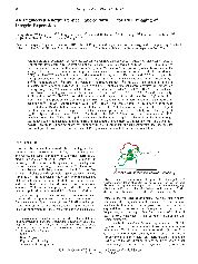
Knottins are small constrained polypeptides that share a common disulfide-bonded framework and a triple-stranded beta-sheet fold. Previously, directed evolution of the Ecballium elateriton trypsin inhibitor (EETI-II) knottin led to the identification of a mutant that bound 10 tumor-specific alpha(v)beta(3) and alpha(v)beta(5) integrin receptors with low nanomolar affinity. The objective of this study was to prepare and evaluate a radiofluorinated version of this knottin (termed 2.5D) for microPET imaging of integrin positive tumors in living subjects. Knottin peptide 2.5D was prepared by solid-phase synthesis and folded in vitro, and its free N-terminal amine was reacted with N-succinimidyl-4-F-18/19-fluorobenzoate (F-18/19-SFB) to produce the fluorinated peptide F-18/19-FB-2.5D. The binding affinities of unlabeled knottin peptide 2.5D and F-19-FB-2.5D to U87MG glioblastoma cells were measured by competition binding assay using I-125-labeled echistalin. It was found that unlabeled 2.5D and F-19-FB-2.5D competed with I-125-echistatin for binding to cell surface integrins with IC50 values of 20.3 +/- 7.3 and 13.2 +/- 5.4 nM, respectively. Radiosynthesis of F-18-FB-2.5D resulted in a product With high specific activity (ca. 100 GBq/mu mol). Next, biodistribution and positron emission tomography (PET) imaging studies were performed to evaluate the in vivo behavior of F-18-FB-2.5D). Approximately 3.7 MBq F-18-FB-2.5D was injected into U87MG tumor-bearing mice via the fail vein. Biodistribution studies demonstrated that F-18-FB-2.5D had moderate tumor uptake at 0.5 h post injection, and coinjection of a large excess of the unlabeled peptidomimetic c(RGDyK) as a blocking agent significantly reduced tumor uptake (1.90 +/- 1.15 vs 0.57 +/- 0.14%ID/g, 70% inhibition, P < 0.05). In vivo microPET imaging showed that F-18-FB-2.5D rapidly accumulated in the tumor and quickly cleared from the blood through the kidneys, allowing excellent turnor-to-normal tissue contrast to be obtained. Collectively, F-18-FB-2.5D allows integrin-specific PET imaging of U87MG turnors with good contrast and further demonstrates that knottins are excellent peptidc scaffolds for development of PET probes with potential for clinical translation.
- 出版日期2009-12